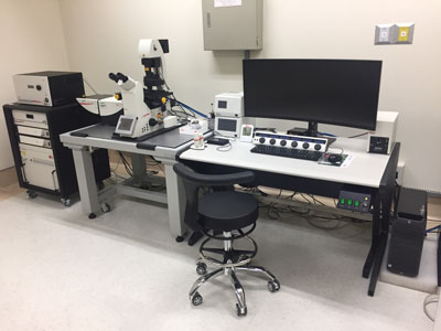
Inverted Laser Scanning Confocal Microscopy (LEICA TCS SP8X STED)
 |
Proposal(Please use the CGMH internet) |
| Publications | |
| Instrument Manager | |
| Name:Chih-Chun Chen | |
| Mei-Ling Huang | |
| Yu Chang | |
| Number:(03)3281200#7706 | |
| 1. | Technical principle | ||||
| 2. | Facility Specifications | ||||
| I. | Type:Leica TCS SP8X STED | ||||
| II. | Light source: | ||||
| i. | Visible laser | ||||
| A. | White light laser system: pulsed laser | ||||
| B. | Super continuous exciting range: 470nm-670nm | ||||
| C. | 1nm adjustable | ||||
| D. | >mW for each wavelength | ||||
| ii. | UV laser | ||||
| A. | 405nm laser | ||||
| B. | 50mW | ||||
| III. | Microscope:LEICA DMi8 Inverted microscope 1set | ||||
| IV. | Objective lens: | ||||
| i. | Plan Fluotar 5x, dry, NA 0.15, WD≧13.7mm | ||||
| ii. | Plan APO 10x, dry, NA 0.40, WD≧2.2mm | ||||
| iii. | Plan APO 20x, NA 0.70, coverglass, WD≧0.59mm | ||||
| iv | Plan APO 40x, oil, NA 1.30, WD≧0.24mm | ||||
| v | Plan APO 63x, oil, NA 1.40, WD≧0.14mm | ||||
| vi | Plan APO 63x, glyc, NA 1.30, CORR, WD≧0.3mm | ||||
| vii | Plan APO 93x, glyc, NA 1.30, motC STED WLL , WD≧0.3mm | ||||
| viii | Plan APO 100x, oil, NA 1.40, STED WHITE , WD≧0.09mm | ||||
| V. | Scanner: | ||||
| i. | for UV/405 nm-VIS-IR-gSTED with 4 independent laser ports | ||||
| ii. | Light gate FLIM technology | ||||
| iii. | High resolution scan mode: | ||||
| A. | Scan field of view(FOV) at least 22 mm, speed 7fps@512x512 in FOV 22mm, 84fps @ 512 x 16 | ||||
| B. | Max. scan resolution 8192 x 8192, 16 bits grey scale, digitalization 12bits or 18 bits. | ||||
| VI. | CCD: | ||||
| i. | 1.4 Mpixel (1392 x 1040) | ||||
| ii. | Exposure time 4us-10min | ||||
| VII. | Image capture format: | ||||
| i. | Bright field | ||||
| ii. | Fluorescence | ||||
| iii. | Reflection | ||||
| iiii. | Normaski differential interference contrast (DIC) | ||||
| VIII. | Build-in Scanner' confocal detector: | ||||
|
Multi-band Spectrophotometer(400-850 nm)with 4PMT tube (2 cooling hybrid GaAsP/APD detectors + 2 cooled PMT detectors) |
|||||
| IX | 3D STED module | ||||
| i. | 592nm STED laser | ||||
| ii. | 660nm STED laser | ||||
| iii. | 775nm STED laser | ||||
| iv. | Resolution dxy~30nm, dz<100nm | ||||
| v. | 3D STED device | ||||
| X | Incubation system | ||||
| XI | Software: | ||||
| i. | Leica Application Suite X | ||||
| ii. | Imaris 3D/4D real time image software | ||||
| iii. | Metamorph image analysis software | ||||