Diagnostic Accuracy of Lateral Neck Radiography for Esophageal Foreign Bodies in Adults
OBJECTIVE. The objective of our study was to evaluate the accuracy of signs on lateral
neck radiography for the assessment of patients with suspected esophageal foreign bodies (FBs).
MATERIALS AND METHODS. This retrospective study was conducted of 235 adult
patients between January 2012 and December 2017. Group 1 was composed of 95 patients
with esophageal FBs, and group 2 was composed of 140 patients without esophageal FBs.
Four signs on lateral neck radiography were recorded in both groups: presence of abnormal
radiopaque density, presence of abnormal air column lucency, loss of cervical lordosis, and
increased prevertebral soft-tissue thickness. The prevertebral thickness was also evaluated in
three groups of patients categorized by patient age: 19–29 years old, 30–59 years old, and 60
years old or older.
RESULTS. The accuracy of the presence of abnormal radiopaque density, presence of
abnormal air column lucency, loss of cervical lordosis, and increased prevertebral soft-tissue
thickness was 84.3%, 66.8%, 54.0%, and 60.9%, respectively. Combined two signs of presence
of radiopaque density with air column lucency provided the highest accuracy, 90.6%. The prevertebral
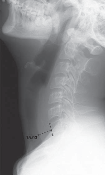
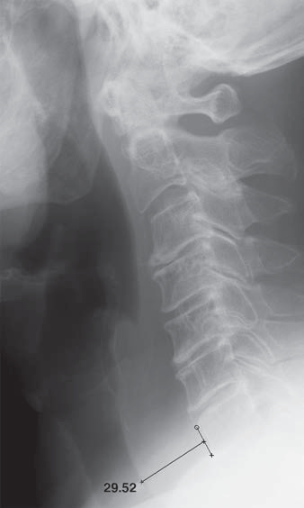
thickness at C6 of group 1 was 14.28 ± 3.19 mm (mean ± SD), and the prevertebral
thickness at C6 of group 2 was 13.34 ± 2.54 mm (p = 0.018).
CONCLUSION. Lateral neck radiography is helpful for the initial evaluation of patients
with suspected esophageal FBs. The presence of radiopaque density or air column lucency
provided the highest practical diagnostic value. Loss of cervical lordosis, as a single diagnostic
sign, did not seem to provide a diagnostic advantage. Despite the variations in values for
the three age groups, clinicians should be alert regarding increased prevertebral thickness at
C6 especially when it is more than 20 mm.
(AJR 2020; 215:1–7, DOI: doi.org/10.2214/AJR.19.21870)